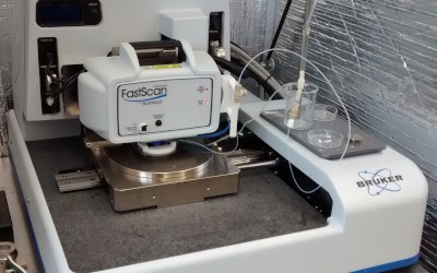
 | Atomic Force Microscopy (AFM)Atomic force microscopy (AFM) is an experimental tool to investigate the topography and mechanical properties of a sample’s surface. Typically, the probe of an AFM is a microscopic, very sharp tip attached to a cantilever. The deflection of the cantilever, caused by the interaction between the tip and the sample surface can be measured via a laser and a photodiode. AFM therefore allows for quantitative topography measurements on the nanometer scale (even sub-nm in z-direction) as well as measurement of, e.g., adhesion force, Young’s modulus or friction. An AFM can be used in different environments, including liquids, and is therefore an excellent tool to investigate biological systems.
|
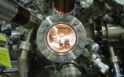 | Ultra High Vacuum LabX-ray photoelectron spectroscopy provides an experimental standard tool for surface analysis. It excels by its very high surface sensitivity, i.e., due to the very small electron mean free paths of electrons with kinetic energies in the range of 10–2000 eV (typically 1-2 nm) only the topmost layers of a sample are probed. Photoelectrons are detected in terms of their distribution of kinetic energies (as a measure of their binding energy) and their distribution of momentum with respect to, e.g., the crystal axes of the sample. In our group we use an
|
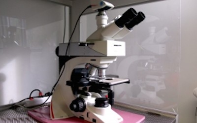 | Optical MicroscopyOptical microscopes use lens systems to magnify structures. Standard microscopes are limited in their resolution to the wavelength of visible light, i.e. a few 100 nm. We use light microscopes, for example, to observe the dewetting of thin films or to study the behaviour of protein layers and vesicles. Depending on the application, we use different microscopes, but primarily the following models
|
|
||||
AG Jacobs
Experimental Physics
Saarland University
Universität Campus
66123 Saarbrücken
Tel: +49 (0) 681 / 302 71777
Fax: +49 (0) 681 / 302 71700
Experimental Physics
Saarland University
Universität Campus
66123 Saarbrücken
Tel: +49 (0) 681 / 302 71777
Fax: +49 (0) 681 / 302 71700
Letters (P.O. Box)
Universität des Saarlandes
Postfach 151 150
Gebäude E2 9
66041 Saarbrücken
Parcels
Universität Campus
Gebäude E2 9, 4. Stock
66123 Saarbrücken
Universität des Saarlandes
Postfach 151 150
Gebäude E2 9
66041 Saarbrücken
Parcels
Universität Campus
Gebäude E2 9, 4. Stock
66123 Saarbrücken
 Deutsche Version
© by AG jacobs 2017-2024 (Saarbrücken)
Legal Notice -- Privacy Policy